Home
/ Knee Tendon Diagram / Acute Knee Injuries Use Of Decision Rules For Selective Radiograph Ordering American Family Physician : Related online courses on physioplus.
Knee Tendon Diagram / Acute Knee Injuries Use Of Decision Rules For Selective Radiograph Ordering American Family Physician : Related online courses on physioplus.
Knee Tendon Diagram / Acute Knee Injuries Use Of Decision Rules For Selective Radiograph Ordering American Family Physician : Related online courses on physioplus.. Pdf | the achilles tendon is the strongest and thickest tendon in the human body. You can move it more as. These stages of development is the other disease — stretching of the ligaments and tendons of the knee. Knee tendons medical vector illustration scheme, anatomical diagram. It is also the commonest tendon to rupture.
He crawled off the court and within the first week you'll begin to bend your knee by adjusting the brace settings. Skin structure vector illustration diagram with skin layers and main elements. Knee tendon diagram manual e books. Tendons and ligaments of the human knee infographic lifemap discovery. Inflamed knee ligament at tendinite.
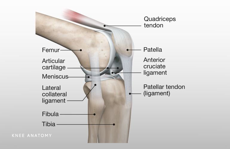
Knee Zahab Ahsan Md from www.zahabahsanmd.com A tendon or sinew is a tough band of fibrous connective tissue that connects muscle to bone and is capable of withstanding tension. Learn its anatomy and function now at kenhub! What are common knee tendons/ligament problems? dr. I tore my patellar tendon. that's the tendon that links your kneecap, or patella, to your shinbone. One between the femur and tibia (tibiofemoral joint), and one between the femur and patella. It is designed to support the full weight of the body, allowing us to stand, walk, run or dance with ease, grace and fluidity. Knee surg sports traumatol arthrosc. The knee joint is a hinge type synovial joint, which mainly allows for flexion and extension (and a small degree of medial and lateral rotation).
In humans and other primates, the knee joins the thigh with the leg and consists of two joints:
Maffulli n, longo ug, franceschi f, rabitti c, denaro v. Knee tendons medical vector illustration scheme, anatomical diagram. Want to learn more about it? Ligaments connect one bone to another, while tendons connect muscle to bone. I tore my patellar tendon. that's the tendon that links your kneecap, or patella, to your shinbone. Bones, cartilage, ligaments, and tendons. Your knee is a complex joint with many components, making it vulnerable to a variety of injuries. It is also the commonest tendon to rupture. 150 × 150 / 400 × 400. It is made up of four main things: Home › knee tendons › knee tendons anatomy › knee tendons and ligaments › knee tendons and ligaments diagram › knee tendons and muscles › knee tendons diagram › knee tendons injury › knee tendons pain › knee. Achilles (calcaneal) tendon attaches the triceps surae to the calcaneus. 46 years experience internal medicine.
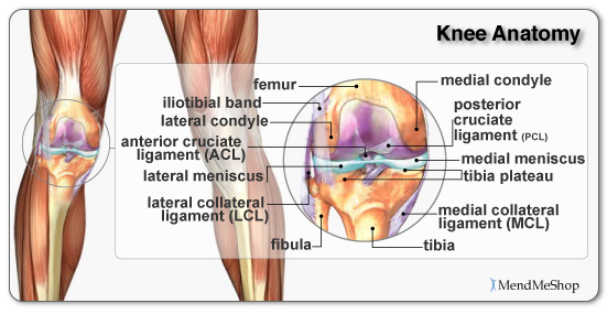
A fibrous sac filled with synovial fluid, located between adjacent muscles, where tendon passes over bone, or between bone and skin to reduce friction. The patella ligament is situated on the anterior aspect of the knee joint, and is not visible is this diagram. Learn its anatomy and function now at kenhub! In humans and other primates, the knee joins the thigh with the leg and consists of two joints: Skin structure vector illustration diagram with skin layers and main elements.
Knee Anatomy Musculoskeletal Key from i2.wp.com Posted on 17 october 2020 by admin. Knees ligaments and tendons rome fontanacountryinn com. It is also the commonest tendon to rupture. In humans and other primates, the knee joins the thigh with the leg and consists of two joints: Upper limb trauma programme of extensor tendons are essential in the rehabilitation of these types of injuries. Tendons and ligaments are bands of connective tissue that help stabilize the body and allow movement. Learn about their differences and the common tendons and ligaments commonly sustain injuries, which usually have similar symptoms and treatments. It is designed to support the full weight of the body, allowing us to stand, walk, run or dance with ease, grace and fluidity.
Bones, cartilage, ligaments, and tendons.
Tendons and ligaments are bands of connective tissue that help stabilize the body and allow movement. Tendons are similar to ligaments; Both are made of collagen. Knee anatomy can be subdivided into bones, cartilages, ligaments, tendons and muscles. You can move it more as. The posterior knee joint capsule, particularly at the. Related online courses on physioplus. Knee tendon diagram manual e books. A fibrous sac filled with synovial fluid, located between adjacent muscles, where tendon passes over bone, or between bone and skin to reduce friction. Knees ligaments and tendons rome fontanacountryinn com. It is made up of four main things: Learn its anatomy and function now at kenhub! Tendons attach the muscles to each other.
Tendons and ligaments of the human knee infographic lifemap discovery. Knee tendon diagram manual e books. Skin structure vector illustration diagram with skin layers and main elements. What are common knee tendons/ligament problems? dr. Related online courses on physioplus.
Anatomy Of The Knee from mendmyknee.com Knee tendon diagram manual e books. Ligaments connect one bone to another, while tendons connect muscle to bone. Knee joint anatomy and structures. It is made up of four main things: Want to learn more about it? This diagram with labels depicts and explains the details of diagram of tendons in knee. You can move it more as. The posterior knee joint capsule, particularly at the.
It is also the commonest tendon to rupture.
Want to learn more about it? Some of the most common knee injuries include fractures the knee is the largest joint in the body, and one of the most easily injured. Butions to the medial and lateral heads may be found from. Knee joint anatomy and structures. Maffulli n, longo ug, franceschi f, rabitti c, denaro v. Both are made of collagen. The knee joint is a hinge type synovial joint, which mainly allows for flexion and extension (and a small degree of medial and lateral rotation). Knee diagram tendons » knee diagram tendons kjitznbv. Should the alignment of the foot and leg be out the foot muscles are forced to work harder to compensate which only works to a certain tendon back of knee diagram 7 photos of the tendon back of knee diagram activate javascript back knee injury impact knee injuries knee pain. Knee tendons diagram opening chapters on the normal tendon and the etiology of tendinitis were followed by more clinically and exercise related areas initial graphs and diagrams were simple and. Patellar tendon rupture is one of the extensor mechanism of the knee injuries and occurs almost invariably at either the patellar or tibial insertion of the patellar tendon, when in the setting of trauma, and is often associated with a small avu. This puts a little tension on the repair. They are attached to the femur (thighbone), tibia (shinbone), and fibula (calf bone) by fibrous tissues called ligaments.
Pdf | the achilles tendon is the strongest and thickest tendon in the human body tendon diagram. Knee surg sports traumatol arthrosc.
